
The Basic Anatomy of the Eye
Although small in size, the eye is quite a complex organ — allowing the brain to interpret what we see, functioning much like a camera.
 Let’s break down the basics of the eye’s anatomy and its many parts that work together to produce clear vision.
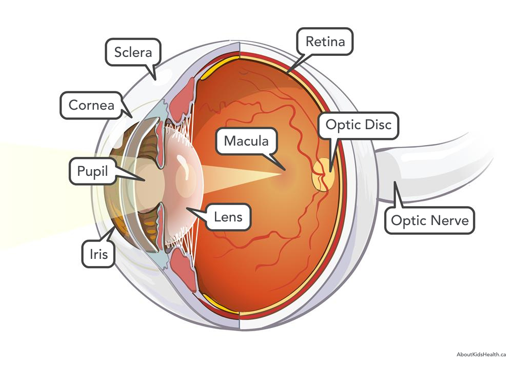
- Cornea: a transparent window at the front of the eye that shields the iris and pupil and transmits the clarity of visuals
- Iris: the colored part of the eye (your eye color), controls how much light enters the eye by changing the size of the pupil
- Lens: acts like a camera lens by focusing light onto the retina at the back of the eye, located behind the pupil
- Macula: the area in the retina that consists of light-sensitive cells.
- Optic Disc: the raised point where the optic nerve enters the retina
- Optic Nerve: millions of nerve fibers that carry visual messages from the retina to the brain.
- Pupil: the black “dot” in the center of the iris, changing its size to allow the amount of light to enter the eye, equivalent to the aperture settings in a camera
- Retina: the nerve layer lining the back of the eye, sensing light and creates electrical impulses that are sent through the optic nerve to the brain
- Sclera: the white outer layer surrounding the eye, protecting the eyeball
Through these features of the eye, visual messages are sent through the eye to the brain to interpret what we see. What the brain interprets is heavily dependent on the transfer of light.
Light passes through the front of the eye (cornea), through the lens, and to the retina. The cornea and lens aid in focusing light rays on the back of the eye (retina). The cells of the retina absorb and convert light into impulses that are sent to the optic nerve and then to the brain.
The eye functions similar to the aperture, shutter, mirror, and lens of a camera. The iris and pupil determine how much light enters the back of the eye (aperture). When darker, the pupils expand, allowing the eye to let in more light. When brighter, the pupils contract to reduce the amount of light that reaches the eye and to avoid over-exposure. The lens of an eye works exactly as it does in a camera. The lens focuses on the details of the visuals. Glasses and contact lenses would be as if changing lenses on a camera, sharpening the details of objects in front of you. Matching the use of a mirror in a camera, the retina sees the visuals upside down and the optic nerve allows the brain to mirror the image right side up.
As you would with an expensive piece of equipment like a camera, you should always protect your eyes. Damage, deterioration, or changes to the anatomy of the eye can lead to vision-related conditions, such as macular degeneration or glaucoma. We recommend routine eye doctor visits to detect any complications or changes to the eye and your vision. Our trained staff will work with you to diagnose any vision problems and suggest the best treatment options and corrective measures for your eyes. Schedule an appointment or visit us at our Galesburg or Monmouth location today!